The Inner Ear
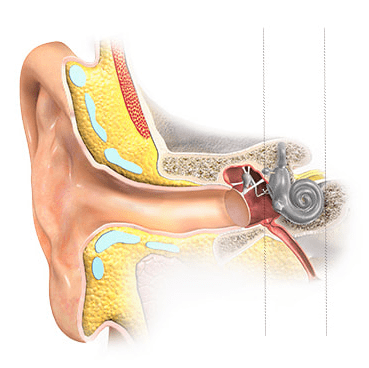
Sound vibrations travel along the ear canal, vibrating the eardrum, and activating the middle-ear bones.
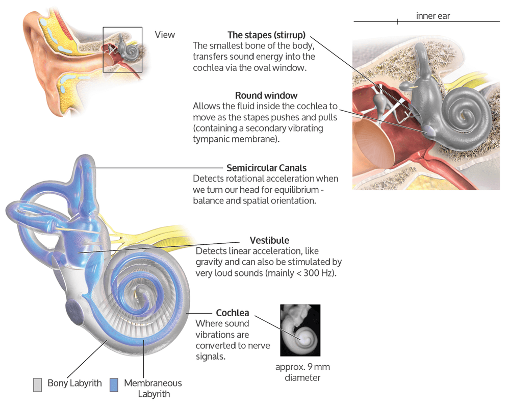
The footplate of the stirrup (stapes) then transfers sound energy through the oval window into the inner ear (the labyrinth), a bony, fluid-filled cochlea - translated from Greek as 'spiral snail shell’. The round window (second tympanic membrane) of the cochlea allows the liquid in the cochlea to slosh around when the stirrup pushes in and out.
Inside the Cochlea
(Cochlea - translated from Greek as 'spiral snail shell')
The cochlea is a liquid-filled ‘shell’ structure about the size of a pea. 9mm at the widest point. Within the cochlea, there are three main channels. In the middle cavity along the basilar membrane are housed 18,000 hair cells. If each hair cell could be brought together, they would fit on the head of a pin. On the top of these hair cells are hair bundles containing 40-80 stereocilia.
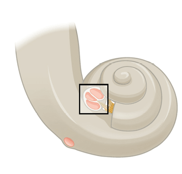
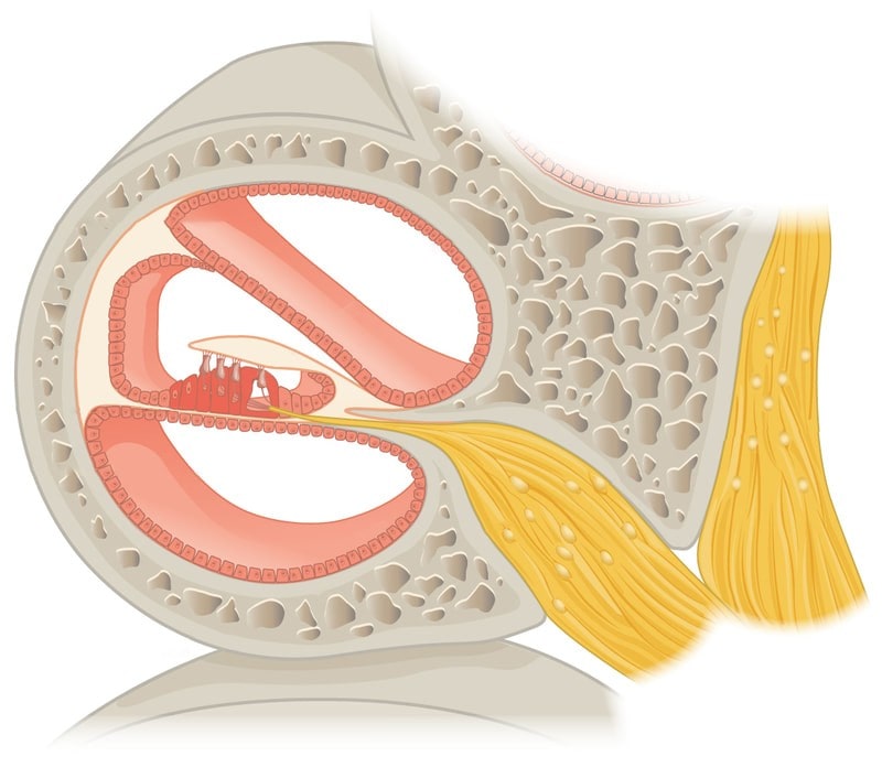
Cochlea cross-section
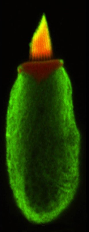
Hair Cell
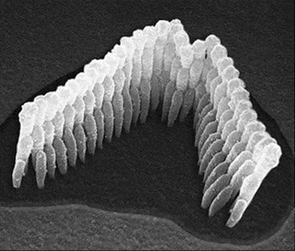
Hair Bundle - Stereocilium
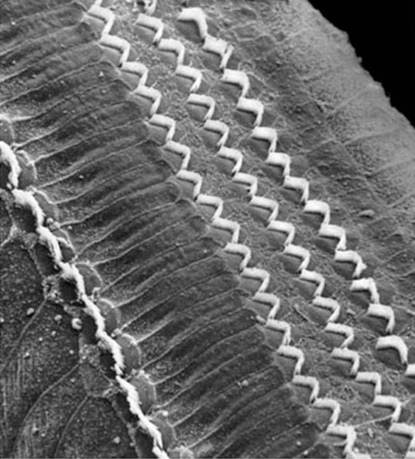
Hair Cells on the Basilar Membrane
Dancing Outer Hair Cell
The three rows of ‘outer hair cells’ on the basilar membrane (above ) amplify sound vibration by a factor of 1000 (much like a 'pre-amp') so that the single row of ‘inner hair cells’ can more effectively process most of the sound vibrations.
When sound vibrations enter the cochlea fluid, the hair bundles 'dance', elongating and contracting, bending and swaying in the inner-ear fluid like ocean seaweed. The video (right) demonstrates the movement of a singular outer hair cell when stimulated by the AC current of music (from Auditory Neuroscience Making sense of sound LINK).
